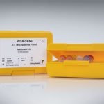
Intended use:
For in vitro diagnostics. This test is an enzyme immunoassay on a nitrocellulose membrane (immunoblot) for the quantitative detection of allergen-specific IgE antibodies in human serum and plasma (citrate).
General information:
The immune system’s task is to protect the organism against pathogenic bacteria, viruses and other microorganisms. The defense reaction protects the organism on first contact with the pathogens, but also provides immunization for recurring exposure. All allergic reactions are preceded by a symptom-free first contact, where specific class E antibodies (IgE antibodies) were already formed. On repeated contact with the triggering allergen these IgE antibodies react with the allergen and cause the release of mediators (usually from mast cells) like histamine, leukotriene, prostaglandin etc., which lead to the allergy symptoms. By detecting the specific IgE antibodies in the serum the triggering allergens can be identified, if there are allergic reactions. But also already existing sensitizations without symptoms can be detected here.
Advantages:
- Economic: allergen panels with up to 20 allergens for effective screening
- Quantitative: real standard curve on each strip (calibrated against the international reference preparation “2nd WHO IRP 75/502 for specific IgE”)
- Customized: customer-specific allergen compositions for flexible and individual diagnostics
- Easy: standardized and convenient performance and evaluation
- Reliable: high concordance of the results for the same allergen with the same sample on different panels and high accuracy of the results in low range results (RAST class 0 – 2)
Test principle:
This test is based on the principles of the immunoblot method. Various allergens are attached to the surface of nitrocellulose membranes in separate lines depending on the configuration of the panel. Allergen-specific IgE antibodies react with the appropriate allergens, if they are present in the patients’ samples. In a second step biotin-conjugated anti-human IgE antibodies bind to the attached antibodies. During a third incubation step the biotin binds to a streptavidin peroxidase conjugate. In a final incubation step the peroxidase turns the colorless substrate tetramethylbenzidine (TMB) into a bluish purple final product. After each individual incubation a washing step follows to remove unbound material. The intensity of the blue color is proportional to the amount of allergen-specific antibodies in the patient’s serum.
The sample is evaluated with RIDA qLine® Scan in combination with the software RIDA qLine® Soft. The color intensities of the allergen bands are quantitatively evaluated on the basis of a standard curve on the membrane to determine the corresponding IU/ml or RAST classes.
New:
Hard- and Software:
- Individual: software evaluation with individually designable report
- Fast: evaluation of 20 strips in one operation with a validated flatbed scanner
- Precise: highly precise detection of the bands and mathematical evaluation of results
- Linkable: connecting to LIS possible
Accessories:
- RIDA® CCD-Inhibitor (en)
- RIDA qLine® Scan
- RIDA qLine® Orbital Shaker (en)
- RIDA qLine® QC-Kit (en)
- RIDA qLine® Incubation Set (en)
| Art. No. | A6142, A6242, A6342, A6442 |
|---|---|
| Test format | Immunoblot Nitrocellulose membrane with 20 allergens on each strip |
| Incubation time | 110 min |
| Shelf life | 24 months |
Dear customers,
we have started to provide the documents for our products in an electronic format. These are the Instructions for Use (IFU), the Safety Data Sheets (SDS) and the Certificate of Analysis (CoA). For batches placed on the market after 01 January 2023, you can find our documents on the eIFU portal eifu.r-biopharm.com.


